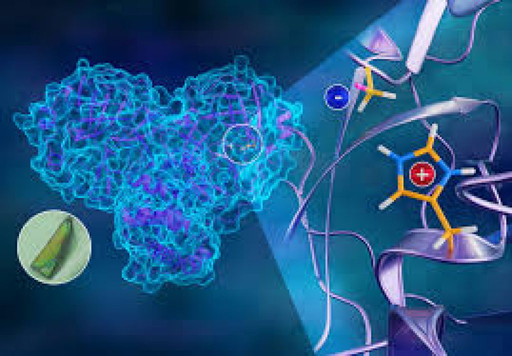
Scientists developed 3D atomic map of novel coronavirus replication mechanism

For the first time, scientists have completed a 3D map that reveals the location of every atom in the molecule of this enzyme. As Covid-19 cases surge again in several countries, this 3D mapping will allow scientists to better understand how the coronavirus behaves, and how it can be stopped.
Daily Current Affairs Quiz 2020
Key-Points
SARS-CoV-2 expresses long chains of proteins. When these chains are broken down and cut into smaller strands, it enables the virus to reproduce.
This task is performed by the main protease. Its structure consists of two identical protein molecules held together by hydrogen bonds.
If a drug can be developed that inhibits or blocks the protease activity, it will prevent the virus from replicating and spreading to other cells in the body.
Researchers used a technique called neutron crystallography. The site containing the amino acids where the protein chains are cut, these experiments revealed, is in an electrically charged reactive state — not in a resting or neutral state, contrary to previously held beliefs.
It is the first time anyone has obtained a neutron structure of a coronavirus protein.
It is also the first time anyone has looked at this class of protease enzymes using neutrons.
The fact that the protein chains are cut at a site that is in an electrically charged reactive state, rather than neutral, was a surprise finding.